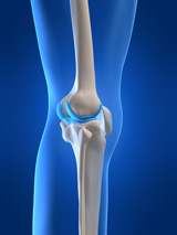
The patella (kneecap) is one of three bones that help form the knee joint, along with the femur (thigh bone) and tibia (shin bone). The kneecap slides up and down a groove on the end of the thigh bone as the knee bends. The kneecap is designed to fit and slide evenly in the center of this groove.
A Patello Femoral Subluxation is when the kneecap is pulled outward towards the outside of the knee. This creates a situation where the kneecap does not properly slide within the normal groove.
A Patello Femoral Dislocation, conversely, is when the kneecap is knocked completely out of position by a direct impact to the knee cap, by a fall, or from a sudden twist of the leg. This can cause intense pain and swelling. Depending on the severity of the kneecap dislocation, the teen or child may experience slight pain, or he or she may experience severe pain around the sides of the kneecap.
Patellar subluxation is more common than a patellar dislocation, but is equally problematic, painful and disabling.
Lateral patellar dislocation can be more common in children. Experts believe that this is because they are young, and there are growth plates that are still maturing. Surgery may be necessary to repair the damage early, especially if the dislocation happens re-occurs after non-surgical treatment options.
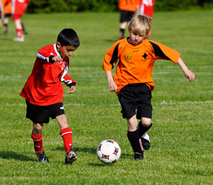
Dislocation typically occurs more in sports that involve knee rotation, or where there can be direct trauma on the knee from football tackles and impact with the ground.
Instability of the kneecap is actually a common problem with young athletes, especially dancers or gymnasts. Non-surgical treatment options sometimes can be linked to recurrent dislocations.
SYMPTOMS
Symptoms of patellar DISLOCATION include:
- Knee appears deformed, bent and cannot be straightened.
- Kneecap appears visibly out of position and toward the outside of the knee.
- Knee swelling, pain/tenderness to touch.
- Kneecap appears loose.
Symptoms of patellar SUBLOXATION include:
- Knee buckles and can no longer support the teen’s bodyweight
- There can be the feeling that the knee catches during range of motion
- The kneecap slips off to the side
- Stiffness in the knee or swelling
- Pain in the kneecap that increases with activity
- Pain when sitting
- Creaking or cracking sounds during movement
Treatment
The pediatric orthopedic specialist examines the knee to determine if it is dislocated. If the knee cap is dislocated, the kneecap will need to be repositioned. Treatment may include surgery or physical therapy to help strengthen the patient’s leg and hip structure or bracing or taping to help support the weakened knee joint. Varsity Orthopedics specializes in conversative surgical options for various knee cap disorders.
SURGERY FOR PATELLAR SUBLUXATION OR DISLOCATION
If the kneecap continues to dislocate multiple times, then patellar stabilization surgery may be recommended. When the kneecap dislocates, the cartilage in the knee can be damaged, which in turn may increase the risk of knee arthritis. Typically, the orthopedic surgeon determines the best surgical approach after looking into the knee with an arthroscope.
Lateral Release: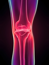
Because the kneecap is being pulled to the outside of the knee, the lateral release surgery cuts the knee joint capsule on the outside of the knee joint, which reduces the tension to the outside and helps re-center the kneecap within the groove on the end of the thigh bone. A lateral release is usually the simplest and most commonly performed surgery to address patellar instability.
Medical Imbrication / Reefing:
While the lateral release surgery method loosens the structures pulling the kneecap outward, medical imbrication is a procedure to tighten the tissue on the inner side of the knee. The pediatric orthopedic surgeon uses an arthroscope to access the joint through a tiny incision to tighten attachments around the knee joint.
MPFL Reconstruction:
The most modern surgery being performed for recurring patellar dislocation is medial patellofemoral ligament (MPFL) reconstruction. The MPFL is the ligament between the end of the thigh bone and the inner side of the kneecap. Typically, when a kneecap dislocates, the MPFL may be torn.
In less severe knee injuries & dislocations, it may be possible to repair the MPFL. This is only true in first-time dislocations that are addressed with immediate surgery. If the patient has experienced multiple dislocations, a new ligament must be created using a ligament or tendon harvested from the patient’s own body, a synthetic ligament, or one harvested from a cadaver, to repair the MPFL. The newly reconstructed MPFL ligament is created, and attached to the thigh bone and kneecap. This holds the kneecap back in proper position.
Bone Realignment:
If the child or teen's knee anatomy is somewhat abnormal, the issue may be caused by a shallow groove on the end of the thigh bone, or abnormal alignment of the lower extremity. The usual surgery is to change the alignment of the thigh bone by shifting the position of the tibial tubercle on the shin bone. The tibial tubercle (the bump at the top of the shin bone) is the attachment point of the patellar tendon. By adjusting the position of the tibial tubercle, the patella is pulled more to the inner side of the knee.
Rehab / Physical Therapy After Surgery:
 Physical therapy is essential to help the patient fully recover from surgery and strengthen their knee and leg structure. It is very important for the patient to maintain their post-surgery rehab plan to help reduce the chances of post-surgery stiffness of the knee. Getting normal strength and mobility recovered after surgery can take months or longer. While dislocations of the patella can still occur after surgery, they are much less common. Most patients are able to resume their pre-injury level of activity without having the risk of dislocating their kneecap.